3D Cultures to study the effects of respiratory infections
In the past 2 years, with the Covid-19 pandemic we have witnessed how a single virus can cripple the economy of nations and impact every day life. Respiratory viral infections, which includes severe acute respiratory syndrome coronaviruses (SARS-CoV) remains among the most common cause of illness and death worldwide. The severe impact it has on health further highlights the importance of developing new tools to study the progression of respiratory virus and explore new therapeutic measures (1).
In the recent years most cell based studies has moved towards 3D cell culture methods. 2D cultures often poorly represent the in-vivo physiology of cells, which leads to non-specific and untranslatable data. In vitro 3D cultures have thus overcome the limitations found in 2D cultures, by precisely mimicking the human tissue structure and function, as well as representing accurate viral tissue growth, kinetics, disease progression and host cell interactions. 3D cultures appears to be the future of viral research, as they permit, efficient screening of anti viral drugs, and testing of efficacy and safety of vaccines and help trace the pathogenicity of new viruses, potentially serving as a monitoring tool for future pandemics (1).
3D cell culture models of coronaviruses
Following the first detection of SARS infection in 2002, there has been an increasing need to find an appropriate model for SARS CoV. Animal models and 2D cultures were not successful in providing a full understanding of the virus. In response researchers moved towards 3D models such as 3D tissue like aggregates produced via co-culturing human bronchias-tracheal mesenchymal cells, and human bronchial epithelial cells in bio reactors. These aggregates were then infected with SARS CoV virus, to investigate the susceptibility and permeability of the cells to the virus. The results were much more significant compared to the 2D cultured counterparts, with respect to structure and function of the cells, viability, and viral protein synthesis; establishing 3D models as an efficient model for the study of coronaviruses (2).
3D culture models to study the pathogenesis of SARS-CoV 2
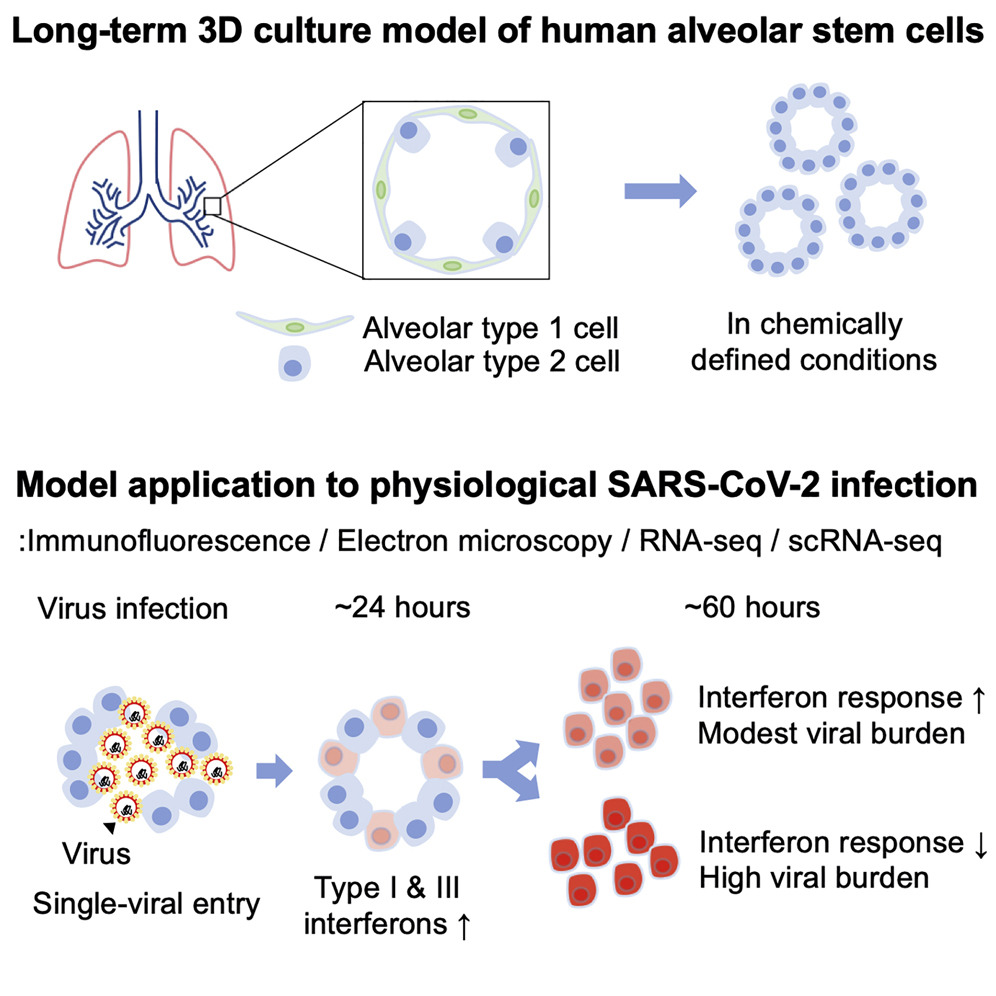
Since the beginning of covid-19 pandemic, much effort has gone in to the study of the virus, and possible treatment options and development of vaccines to combat the effects of SARS-CoV 2. To this effect human pluripotent stem cell-derived lung and colonic organoids (hPSC-LOs and hPSC-COs) were developed to better understand the disease progression of COVID-19, as well as investigate possible drugs effective at preventing virus entry into host cells (3). Additionally investigative teams in Germany have also developed human intestinal organoids derived from pluripotent stem cells (PSC-HIOs) to study SARS-CoV-2 pathogenesis and its inhibition by Remdesivir, the first FDA-approved drug to treat COVID-19 (4). Furthermore 3D model of human alveoli has also been developed to study the entry of SARS CoV in to host cells and the resultant inflammatory response (5). iPSCs-derived 3D human brain organoids were successfully developed as a model system to test the neurotoxic effects of SARS-CoV-2 (6). These models collectively demonstrate the capability of 3D cultures to be a reliable in vitro human model to study effects of SARS-CoV-2 infection and provide a relevant, modifiable platform for drug screens to identify potential COVID-19 therapeutics.
References
1. Harb, A.; Fakhreddine, M.; Zaraket, H.; Saleh, F.A. Three-Dimensional Cell Culture Models to Study Respiratory Virus Infections Including COVID-19. Biomimetics 2022,7,3.
2. Suderman,M.T.;McCarthy,M.;Mossell,E.;Watts,D.M.;Peters,C.J.;Shope,R.;Goodwin,T.J.Three-DimensionalHumanBronchial- Tracheal Epithelial Tissue-Like Assemblies (TLAs) as Hosts for Severe Acute Respiratory Syndrome (SARS)-CoV Infection; NASA Tech. Paper; NASA Johnson Space Center: Houston, TX, USA, 2006.
3. Rijsbergen, L.C.; Van Dijk, L.; Engel, M.; De Vries, R.D.; De Swart, R.L. In Vitro Modelling of Respiratory Virus Infections in Human Airway Epithelial Cells- A Systematic Review. Front. Immunol. 2021, 12, 683002.
4. Krüger,J.;Groß,R.;Conzelmann,C.;Müller,J.A.;Koepke,L.;Sparrer,K.M.;Weil,T.;Schütz,D.;Seufferlein,T.;Barth,T.F.;etal.Drug Inhibition of SARS-CoV-2 Replication in Human Pluripotent Stem Cell–Derived Intestinal Organoids. Cell. Mol. Gastroenterol. Hepatol. 2021, 11, 935–948
5. Mulay,A.;Konda,B.;Garcia,G.;Yao,C.;Beil,S.;Villalba,J.M.;Koziol,C.;Sen,C.;Purkayastha,A.;Kolls,J.K.;etal.SARS-CoV-2 infection of primary human lung epithelium for COVID-19 modeling and drug discovery. Cell Rep. 2021, 35, 109055.
6. Ramani, A.; Müller, L.; Ostermann, P.N.; Gabriel, E.; Abida-Islam, P.; Müller-Schiffmann, A.; Mariappan, A.; Goureau, O.; Gruell, H.; Walker, A.; et al. SARS -CoV-2 targets neurons of 3D human brain organoids. EMBO J. 2020, 39, e106230.
7. Jeonghwan Youk, Taewoo Kim, Kelly V. Evans, Young-Il Jeong, Yongsuk Hur, Seon Pyo Hong, Je Hyoung Kim, Kijong Yi, Su Yeon Kim, Kwon Joong Na, Thomas Bleazard, Ho Min Kim, Mick Fellows, Krishnaa T. Mahbubani, Kourosh Saeb-Parsy, Seon Young Kim, Young Tae Kim, Gou Young Koh, Byeong-Sun Choi, Young Seok Ju, Joo-Hyeon Lee,
8. Three-Dimensional Human Alveolar Stem Cell Culture Models Reveal Infection Response to SARS-CoV-2, Cell Stem Cell, Volume 27, Issue 6, 2020, Pages 905-919.e10.


