How to Generate Multicellular Spheroids
Since the introduction of monolayer cell cultures, cell culture methods have advanced exponentially in the past decade, moving from traditional 2D cell culture methods to 3D cultures. While 2D cultures are simple and allows for cell proliferation and differentiation, they are unable to recapitulate the complex microenvironment found in the body and, as a result, they lose tissue specific properties which are vital to obtain an accurate representation of in vivo biological situation in the lab.
In contrast, 3D cultures are better equipped to replicate in vivo cell-cell, and cell- extracellular matrix interactions thus providing more realistic microenvironment for in vivo-like cellular behaviour (1).
What are multicellular spheroids (MCTS)?
Multicellular spheroids are the result of spontaneous aggregation of cells. These clusters of cells can form in the presence of cadherins. Cadherins are adhesion molecules that are necessary for forming adherent junctions that causes cells to stick to each other. They are type -1 transmembrane proteins, and depend on calcium ions for proper function (2).
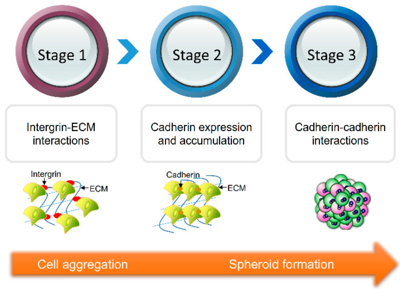
Stages of multicellular spheroid formation
Stage 1
In the first stage, the extracellular matrix containing few RGD motifs operates as long link head. In response to these motifs, single cells in suspension form a loosely packed aggregates under the action of integrins..
Stage 2
In the second stage, the expression and accumulation of epithelial cadherins increase leading to prolonged phase of compaction.
Stage 3
In the third and final stage, the loosely aggregated cells form dense spheroids via cadherin-cadherin binding. Additional external mechanical stimuli conveyed to actin filaments by integrins further encourage spheroid self-assembly.
MCSs have become a useful 3D model for tumor research, anti-tumor drug screening, drug toxicity assays and tissue engineering. Apart from their diverse use in many research fields, they help to reduce operating cost and ethical concerns derived from the use of animal models (3-6).
References
1. Li, A.; Yang, P.M. Overexpression of miR-21-5p in colorectal cancer cells promotes self-assembly of E-cadherin-dependent multicellular tumor spheroids. Tissue Cell 2020, 65, 101365.
2. Lin, R.Z.; Chou, L.F.; Chien, C.C.; Chang, H.Y. Dynamic analysis of hepatoma spheroid formation: Roles of E-cadherin and beta1-integrin. Cell Tissue Res. 2006, 324, 411–422.
3. Jang, M.; Koh, I.; Lee, S.J.; Cheong, J.H.; Kim, P. Droplet-based microtumor model to assess cell-ECM interactions and drug resistance of gastric cancer cells. Sci. Rep. 2017, 7, 41541.
4. Ko, J.; Ahn, J.; Kim, S.; Lee, Y.; Lee, J.; Park, D.; Jeon, N.L. Tumor spheroid-on-a-chip: A standardized microfluidic culture platform for investigating tumor angiogenesis. Lab Chip 2019, 19, 2822–2833
5. Liu, Z.; Vunjak-Novakovic, G. Modeling tumor microenvironments using custom-designed biomaterial scaffolds. Curr. Opin. Chem. Eng. 2016, 11, 94–105.
6. Shen, H.; Cai, S.; Wu, C.; Yang, W.; Yu, H.; Liu, L. Recent Advances in Three-Dimensional Multicellular Spheroid Culture and Future Development. Micromachines 2021, 12, 96.