Is It The Right Time to Advance from 2D – 3D Cell Culture
Since the early 1900s, the two-dimensional (2D) cell culture method is used to culture cells and grow them in favorable artificial conditions (1) thereby playing a vital role in biomedical research (2). In line with the limitations of 2D cell cultures, another method known as three-dimensional (3D) cell culture has been developed and proven to be an improved technique for research application in cell morphology, proliferation, stimuli response, drug metabolism, and protein synthesis, among others (3).
Therefore, 3D cell cultures have applications in many fields such as cancer research, stem cell research, drug discovery, and research on other types of diseases, making it a versatile choice for scientists. While 2D cell culture has traditionally been a very stable method of culturing cells in a standardized and rather simple way, 3D cell culture outperforms 2D cell culture methods in terms of the ability to reproduce the in vivo situation, even though 3D cell culture methods are still in their early stages.
The conventional 2D cell culture approach
2D cell culture methods involve the use of an artificial surface such as the bottom of a petri dish or flask to nourish and grow cells and represent well-established techniques. Despite it being an inexpensive and easy method to use, 2D cell culture comes with certain limitations.
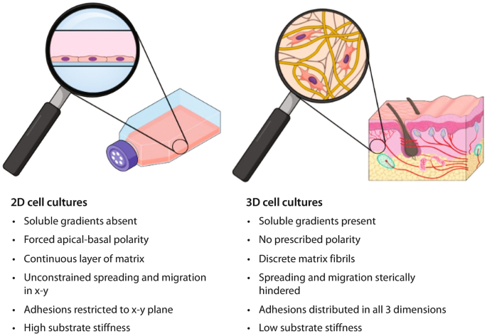
In our body, the cells do not grow in a 2D environment, so 2D cell culture systems lack accuracy in simulating the conditions the cells would encounter in vivo, failing to reproduce normal cell physiology (4). A 2D cell culture system also fails to mimic the diffusion of oxygen and other molecules in the in vivo environment of the cells. Cells being grown as a monolayer are equally exposed to the culture medium with drugs, growth factors, or additives in it.
This is not the case for cells in our bodies. Additionally, 2D cell system cells do not offer an accurate drug-response prediction rate. Because 2D cell cultures are relatively inflexible and only a limited range of cellular behaviors can be tested, 3D cell culture has been gaining attention in recent years.
The new dimension of cell culture
One of the major differences between the 3D and 2D cell cultures is the use of a suitable ‘scaffold’ in 3D cell cultures. 3D scaffolds can act as extracellular matrix (ECM) and are usually made of hydrogel, collagen, or polystyrene acting as physical support for the cells. 3D cell culture approaches reduce the reliance on animal models by providing more accurate simulations of cell natural environment increasing the accuracy, safety, and sustainability of cell culture analyses.
All these features of a 3D cell culture have gained attention within tumor cell biology research because of their ability to replicate the in vivo environment of a tumor cell (5). For example, recent success in treating melanoma skin cancer has encouraged researchers to model melanoma tumor cells in 3D spheroid cell culture to target molecular mechanisms that are involved in resistance in current immunotherapy treatments (6). 3D cell culturing methods have the potential to unlock the answers researchers have been unable to uncover through the use of 2D cell culture techniques. Despite the challenge of it being a more expensive and labor-intensive technique, 3D cell culture is here to stay and to open up new possibilities for analyses in a wide range of areas.
References
1. Barcellos-Hoff MH, Aggeler J, Ram TG, Bissell MJ. Functional differentiation and alveolar morphogenesis of primary mammary cultures on reconstituted basement membrane. Development. 1989 Feb;105(2):223-35. doi: 10.1242/dev.105.2.223. PMID: 2806122; PMCID: PMC2948482.
2. Costa EC, Moreira AF, de Melo-Diogo D, Gaspar VM, Carvalho MP, Correia IJ. 3D tumor spheroids: an overview on the tools and techniques used for their analysis. Biotechnol Adv. 2016 Dec;34(8):1427-1441. doi: 10.1016/j.biotechadv.2016.11.002. Epub 2016 Nov 11. PMID: 27845258.
3. Eltom, Abdalla & Zhong, Gaoyan & Ameen, Muhammad. (2019). Scaffold Techniques and Designs in Tissue Engineering Functions and Purposes: A Review. Advances in Materials Science and Engineering. 2019. 1-13. 10.1155/2019/3429527.
4. Edmondson R, Broglie JJ, Adcock AF, Yang L. Three-dimensional cell culture systems and their applications in drug discovery and cell-based biosensors. Assay Drug Dev Technol. 2014 May;12(4):207-18. doi: 10.1089/adt.2014.573. PMID: 24831787; PMCID: PMC4026212.
5. Holmes TD, El-Sherbiny YM, Davison A, Clough SL, Blair GE, Cook GP. A human NK cell activation/inhibition threshold allows small changes in the target cell surface phenotype to dramatically alter susceptibility to NK cells. J Immunol. 2011 Feb 1;186(3):1538-45. doi: 10.4049/jimmunol.1000951. Epub 2010 Dec 29. PMID: 21191066.
6. Müller I, Kulms D. A 3D Organotypic Melanoma Spheroid Skin Model. J Vis Exp. 2018 May 18;(135):57500. doi: 10.3791/57500. PMID: 29863656; PMCID: PMC6101273