Forming Compact Spheroids
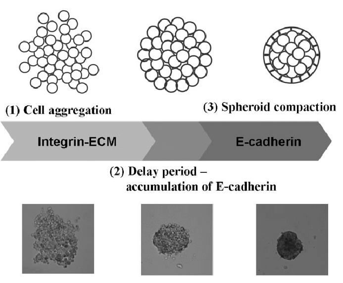
Spheroids, which are self, assembled cellular aggregates, as a representative 3D model of solid tumors is largely dependent on the uniformity of its size and shape. Uniform spheroids yield reproducible results in drug screens as well as obtaining meaningful findings in tumor pathophysiological research. Additionally uniform spheroids, aids in quantification of treatment options, and estimate the effect of treatment uncertainty on the results.
Compact spheroids are often found resistant to drug intervention when compared with aggregated cells, and smaller spheroids are vulnerable to chemotherapy and radiotherapy. Larger multicellular spheroids (MCTs) are composed of tight cell-cell adhesions preventing drug penetration and contain hypoxic cells increasing resistance to therapeutic intervention. Finally formation of uniform, compact spheroids increases its potential use in high throughput drug screens, thus making them preferable model in medical research (1-6).
Methods to form compact MCTs
There have been several methods developed to ensure formation of MCTs. One of which includes the addition of a reconstituted basement membrane in the culture media to facilitate uniform morphology (7-9). These additives include Matrigel, rBM, Geltrex® and collagen. To this effect, Matrigel promoted the formation of uniform and compact spheroids of breast cancer cell line MDA-MB-231 which typically contain low levels of E-cadherin necessary for cell-cell tight junctions. Other breast cancer cell lines cultured in the presence of 2.5% rBM also indicated compact spheroids of uniform size and shape (3).
Another method proposed to encourage the formation of uniform spheroids is micrfabrication of microwells. The microwells are fabricated using 3D printing technology and soft lithography. This is a fairly easy method, offering simple control of well size and compatible with most methods of culturing. Synthetic polymers such as PMMA, polystyrene, PDMS, and PEGDMA are employed are fabrication. Other natural polymers such as collagen, gelatin and agarose are also used. The microwell dimensions spatially confine the cells, leading to formation of spheroids of controlled size and shape (6,10).
Other techniques employed to generate uniform spheroids includes microprinting of MCTs using the aqueous two-phase system (ATPS), methods involving shear flow under orbital shaking of media. In the latter, careful monitoring of the culture and shaking times regulate the size of spheroids. To obtain larger spheroids culture and shaking times are extended (10).
References
1. Bufa FM, West C, Byrne K, Moore JV, Nahum AE. Radiation response and cure rate of human colon adenocarcinoma spheroids of diferent size. Int J Radiat Oncol Biol Phys. 2001;49(4):1109–18.
2. Horas JA, Olguin OR, Rizzotto MG. On the surviving fraction in irradiated multicellular tumour spheroids: calculation of overall radiosensitivity parameters, infuence of hypoxia and volume efects. Phys Med Biol. 2005;50(8):1689–701.
3. Weber W, Weber J, Senekowitsch-Schmidtke R. Therapeutic efect of m-[’3’I]- and m-[’251] Iodobenzylguanidine on neuroblastoma multicellular tumor spheroids of diferent sizes. Cancer Res. 1996;56(23):5428–34.
4. Silvio D, Nunzia A, Diego C, Robert I, Eleonora T, Raoul AD, et al. Induction of hypoxia and necrosis in multicellular tumor spheroids is associated with resistance to chemotherapy treatment. Oncotarget. 2017;8(1):1725–36.
5. Gencoglu MF, Barney LE, Hall CL, Brooks EA, Schwartz AD, Daniel CC, et al. Comparative study of multicellular tumor spheroid formation methods and implications for drug screening. ACS Biomater Sci Eng. 2018;4(2):410–20.
6. Lee JM, Park DY, Yang L, Kim EJ, Ahrberg CD, Lee KB, et al. Generation of uniform-sized multicellular tumor spheroids using hydrogel microwells for advanced drug screening. Sci Rep. 2018;8(1):17145.
7. Badea MA, Balas M, Hermenean A, Ciceu A, Herman H, Daniela I, et al. Infuence of matrigel on single- and multiple-spheroid cultures in breast cancer research. SLAS Discov. 2019;24(5):563–78.
8. Beckingham LJ, Todorovic M, Velasquez JT, Vial ML, Chen M, Jenny AKE, et al. Three-dimensional cell culture can be regulated by vibration: low-frequency vibration increases the size of olfactory ensheathing cell spheroids. J Biol Eng. 2019;13:41.
9. Leung BM, Lesher-Perez SC, Matsuoka T, Moraes C, Takayama S. Media additives to promote spheroid circularity and compactness in hanging drop platform. Biomater Sci. 2015;3(2):336–44.
10. Han, S.J., Kwon, S. & Kim, K.S. Challenges of applying multicellular tumor spheroids in preclinical phase. Cancer Cell Int 21, 152 (2021).
11. Lin, Ruei-Zeng & Lin, Ruei-Zhen & Chang, Hwan-You. Recent advances in three-dimensional multicellular spheroid culture for biomedical research. Biotechnology journal. (2008). 3. 1172-84.