AN ERA OF 3D CULTURES : A BREIF OVERVIEW ON SPHEROIDS
Wondering what these tiny clumps of cells are? let’s have a look.
WHAT ARE SPHEROIDS?
Spheroids are three-dimensional (3D) cell aggregate to better stimulate a in-vivo like environment as compared to a two-dimensional model. Compared to other 3D cell culture models, spheroids have gained an increasing attention due to its relevance in tumor biology, lower processing costs, a better alternative to animal models, higher reproducibility, advanced imaging techniques and ease of integration to higher-throughput screening. (1)
CREATE YOUR OWN SPHEROID
Spheroids can be grown with few different methods. One of them is to use a low cell adhesion plate, typically a 96 well plates such as faCellitate BIOFLOAT™ 96-Well Plate, to mass produce spheroid cultures where the aggregates form in round bottom of cell plates. Spheroids can also be cultured using hanging drop method involving forming of cell aggregates in droplets that hang from the surface of cell plates. Yet, another method used includes the use of rotating wall vessels, which spins the cultures, promoting the formation of cell aggregates in layers. (1)
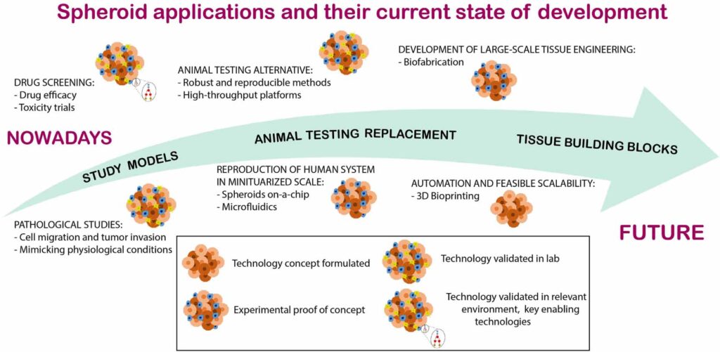
APPLICATIONS AND ADVANCEMENTS
The 3D culture systems not only provide the spatial cell–cell interactions and cell–ECM interactions in monoculture for studying cell behaviors that mimic in vivo conditions, but also provide an opportunity for the co-culture of multiple types of cells to more closely mimic the in vivo conditions. The use of in vitro cell models in drug screening trials has advantages such as the possibility of using smaller sample quantities, added to greater process simplicity and reproducibility when compared to animal trials (2). Tumor spheroids appear as a promising model for the selection of new drug candidates for cancer therapy, since they are able to recapitulate, among other characteristics, the 3D architecture of the microenvironment where tumor cells grow cultures to mimic natural cellular responses in vitro (3). Spheroids and organoids are also used as models for developmental studies and to elucidate mechanisms related to different types of pathologies due to their ability to mimic physiological conditions of specific tissues and organs. All in all, recent developments in 3D culture system have shown to be the more likely preferred method over 2D culture systems.
References:
1. Jiang Y, Pjesivac-Grbovic J, Cantrell C, Freyer JP (December 2005). “A multiscale model for avascular tumor growth”. Biophysical Journal.
2. Van Vliet E 2014 Current standing and future prospects for the technologies proposed to transform toxicity testing in the 21st century ALTEX
3. Lee J, Lilly D, Doty C, Podsiadlo P and Kotov N 2009 In vitro toxicity testing of nanoparticles in 3D cell culture