Types of three-dimensional cultures for drug screening
The rather low success rates in new drug development and the increased costs suggest that alternative and robust strategies are required to improve early drug discovery.
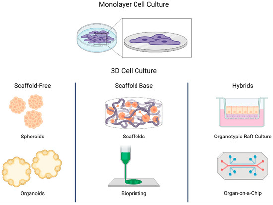
Two-dimensional (2D) monolayer cultures do not provide a true in vitro representation of disease and the introduction of more relevant cell models and high-content imaging techniques is expected to increase the success of drug development processes. This article provides an overview of different three-dimensional (3D) cell culture assays and their implications for drug screening.
1. Co-culture: Cocultures of different cell types are commonly used to understand the functional interactions between different cells and can be performed both in 2D and 3D. The automation of co-culture assays for drug screening is relatively simple but requires large-scale standardization of the procedures. Variations in the assays increase with the number of cell types that are co-cultured (1).
2. Spheroid: Spheroids are 3D cell aggregates that self-assemble into sphere-like structures and promote cell–to–cell interactions. They can mimic tissues and microtumors more faithfully than 2D cultures and are generally cultured in an anchorage-independent context from cell lines grown in 2D cell culture. Automation for drug screening can be readily achieved depending on the cell type and the plates that are used for spheroid formation (2,3).
3. Microtissue: Microtissues are a kind of microscopic tissue formed by the aggregation of primary cells upon cell–cell, and cell–ECM interaction. The microstructure of these 3D structures is similar to that of natural tissues. The main challenge towards automation for drug screening is represented by ECM preparation. In general, gel-embedded cultures are more challenging than gel-coated plates.
4. Organoid: An organoid is a simple tissue-engineered model produced in vitro in which cells self-organize forming miniaturized organs. These 3D structures mimic the key functional, structural, and biological complexity of human organs. Patient-derived primary cells isolated from specific tissues can be used to generate organoids in vitro to improve the relevance of cell culture assays. However, like microtissues, handling ECM preparation is challenging for automation equipment for drug screening. Slow growth, high reagent cost, and time-intensive procedures further limit automation (4).
References:
1. Wang PC, Takezawa T. Reconstruction of renal glomerular tissue using collagen vitrigel scaffold. J Biosci Bioeng. 2005 Jun;99(6):529-40. doi: 10.1263/jbb.99.529. PMID: 16233828.
2. Herter S, Morra L, Schlenker R, Sulcova J, Fahrni L, Waldhauer I, Lehmann S, Reisländer T, Agarkova I, Kelm JM, Klein C, Umana P, Bacac M. A novel three-dimensional heterotypic spheroid model for the assessment of the activity of cancer immunotherapy agents. Cancer Immunol Immunother. 2017 Jan;66(1):129-140. doi: 10.1007/s00262-016-1927-1. Epub 2016 Nov 17. PMID: 27858101; PMCID: PMC5222939.
3. Kang JH, Gimble JM, Kaplan DL. In vitro 3D model for human vascularized adipose tissue. Tissue Eng Part A. 2009 Aug;15(8):2227-36. doi: 10.1089/ten.tea.2008.0469. PMID: 19207036; PMCID: PMC2792112.
4. Calandrini C, Drost J. Normal and tumor-derived organoids as a drug screening platform for tumor-specific drug vulnerabilities. STAR Protoc. 2022 Jan 10;3(1):101079. doi: 10.1016/j.xpro.2021.101079. PMID: 35036959; PMCID: PMC8752949.