Therapeutic Efficacy Of Multicellular Tumor Spheroids
Despite the much advancement seen in tumor related research and drug discovery, cancer research still experiences a significant amount of challenges due the complex behaviour of tumor cells. Multicellular tumor spheroids (MCTs) bear remarkable similarities to in vivo solid tumors and are thus presented as a viable candidate to examine and predict response to chemotherapy and radiotherapy. However the efficiency of MCTs as a reproducible model relies on the availability of appropriate evaluation methods.
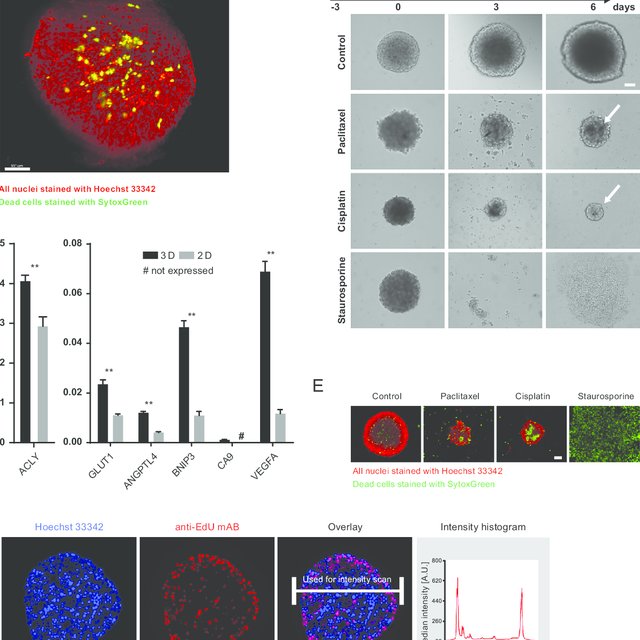
Changes in shape and volume of MCTs
Drug and therapeutic efficacy of MCTs is firstly evaluated by changes in the shape and volume of the spheroids as a direct response to therapeutic intervention. With continuous drug treatment, these morphological changes are followed by alterations in cell-cell and cell – extracellular matrix (ECM) connections, leading to disruptions in cellular aggregations. As the cytotoxicity increases, the outer layer cells begins to fall away, ultimately resulting in the collapse of spherical shape. As treatment progresses, the volume of the spheroid also decreases in a dose dependent fashion (1-2).
Role of compactness and size in evaluating drug efficacy
Therapeutic efficacy of MCTs is also determined by the compactness of the spheroids. Compact spheroids exhibit higher drug resistance compared the loosely aggregated spheroids. This is primarily due to high content of ECM present in compact spheroids, hindering drug delivery to the core of the spheroid. Thus in order to obtain a reliable drug efficacy result the MCTs must be of a uniformly shaped and compact aggregate (3).
The size of the spheroid also plays a role in its drug response. Smaller spheroids are found more sensitive to therapeutic treatment, whereas larger spheroids indicate increased drug resistance (4-7). For example, doxorubicin (DOX), a chemotherapeutic drug is able to easily penetrate smaller MCF-7 spheroids, but is only able to affect the outer layer of the larger spheroids (8). Therefore, much attention is now being devoted to developing MCTS that are uniform, compact and homogenous in size, in order to be standardized as an appropriate solid tumor model that can be used on a high throughout platform.
References
1. Grandis RA, dos Santos PWS, de Oliveira KM, Machado ART, Aissa AF, Alzir AB, et al. Novel lawsone-containing ruthenium (II) complexes: Synthesis, characterization and anticancer activity on 2D and 3D spheroid models of prostate cancer cells. Bioorg Chem. 2019;85:455–68.
2. Dubois C, Dufour R, Daumar P, Aubel C, Szczepaniak C, Christelle B, et al. Development and cytotoxic response of two proliferative MDAMB-231 and non-proliferative SUM1315 three-dimensional cell culture models of triple-negative basal-like breast cancer cell lines. Oncotarget. 2017;8(56):95316–31.
3. Raghavan S, Mehta P, Horst EN, Ward MR, Rowley KR, Mehta G. Comparative analysis of tumor spheroid generation techniques for diferential in vitro drug toxicity. Oncotarget. 2016;7(13):16948–61.
4. Bufa FM, West C, Byrne K, Moore JV, Nahum AE. Radiation response and cure rate of human colon adenocarcinoma spheroids of diferent size. Int J Radiat Oncol Biol Phys. 2001;49(4):1109–18.
5. Horas JA, Olguin OR, Rizzotto MG. On the surviving fraction in irradiated multicellular tumour spheroids: calculation of overall radiosensitivity parameters, infuence of hypoxia and volume efects. Phys Med Biol. 2005;50(8):1689–701.
6. Silvio D, Nunzia A, Diego C, Robert I, Eleonora T, Raoul AD, et al. Induction of hypoxia and necrosis in multicellular tumor spheroids is associated with resistance to chemotherapy treatment. Oncotarget. 2017;8(1):1725–36.
7. Gaze MN, Mairs RJ, Boyack SM, Wheldon TE, Barrett A. 1’I-meta-iodobenzylguanidine therapy in neuroblastoma spheroids of diferent size. Br J Cancer. 1992;66(6):1048–52.
8. Deisboeck TS, Berens ME, Kansal AR, Torquato S, Stemmer-Rachamimov AO, Chiocca EA. Pattern of self-organization in tumour systems: complex growth dynamics in a novel brain tumour spheroid model. Cell Prolif. 2001;34(2):115–34