Introduction to fluorescence microscopy: Principle and types
Fluorescence is the property of some atoms and molecules to absorb radiation at a particular wavelength and subsequently emit light at a longer wavelength upon excitation of the atoms. A fluorescence microscope is a powerful optical tool that works based on the principle of fluorescence. German physicists Otto Heimstaedt and Heinrich Lehmann developed the first fluorescence microscopes in the early 1900s as a spin-off of the ultraviolet instrument. They employed these microscopes to observe autofluorescence in bacteria, animal, and plant tissues (1).
Fluorescent microscopy has enabled scientists to obtain images with a better resolution using fluorophore tags. Usually, the specimens of interest need to be stained with fluorescent dyes called fluorophores to be viewed under a fluorescent microscope because they do not fluoresce naturally. Some commonly used fluorescent dyes are: 4´,6-diamidino-2-phenylindole, acridine orange, auramine-rhodamine, and Alexa Fluors.
Principle
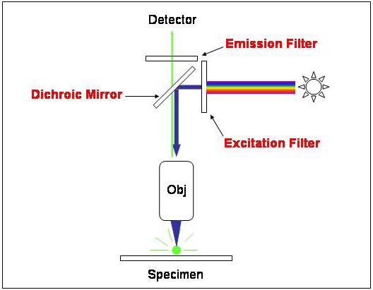
The fundamental principle behind fluorescent microscopes is the use of fluorescent dyes/substances, also called fluorophores, for labeling the cellular structures of interest. Light with high energy and short wavelength is generated from mercury vapor arc lamps and passes through an excitation filter which further allows only the short wavelength of light to pass through, removing all other non-specific wavelengths of light. A dichroic filter is used to reflect the filtered light which falls on the fluorophore-labeled sample.
The fluorophore absorbs shorter wavelength radiations and emits radiations of longer wavelengths that pass through the emission filter. The emission filter allows the desired wavelength radiation to reach the detector, blocking any other residual excitation light. This way, the fluorescent microscope generates coloured images of the fluorophore-labeled samples against a dark background (2).
Types of fluorescence microscopy
1. Epifluorescence microscopy: The term “epi” comes from Greek and means “same”. In epifluorescence microscopes, the fluorescence of the specimen resulting in emitted light is focused on the detector using the same objective used for excitation.
2. Confocal microscopy: The confocal microscope uses spatial filtering that blocks out-of-focus light for a high-resolution 3D image of the sample.
3. Multiphoton microscopy: Multiphoton microscopy is used to visualize cellular and subcellular events within living tissue. It is the most convenient approach for deeper penetration and minimal photobleaching (3).
References
1. Sluder G, Nordberg JJ. Microscope basics. Methods Cell Biol. 2013;114:1-10. doi: 10.1016/B978-0-12-407761-4.00001-4. PMID: 23931500.
2. Sanderson MJ, Smith I, Parker I, Bootman MD. Fluorescence microscopy. Cold Spring Harb Protoc. 2014 Oct 1;2014(10):pdb.top071795. doi: 10.1101/pdb.top071795. PMID: 25275114; PMCID: PMC4711767.
3. Jensen EC. Types of imaging, Part 2: an overview of fluorescence microscopy. Anat Rec (Hoboken). 2012 Oct;295(10):1621-7. doi: 10.1002/ar.22548. Epub 2012 Sep 3. PMID: 22941879.