How to create three-dimensional cell culture models
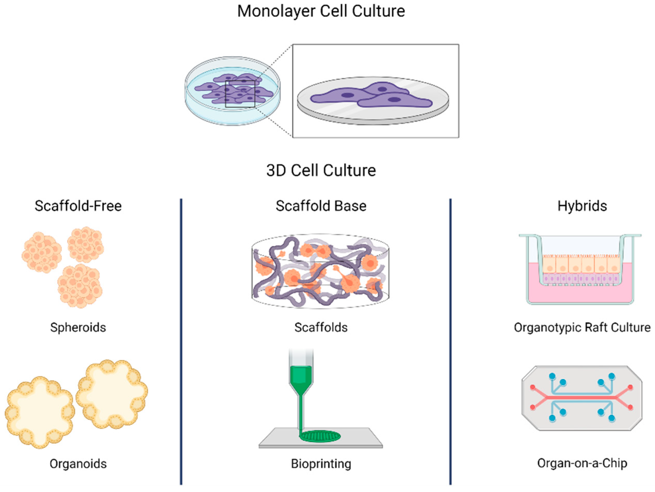
A three-dimensional (3D) cell culture is an artificially created environment in which cells grow or interact with their surroundings in all three dimensions. This cell culture is becoming increasingly popular to replace the traditional two-dimensional (2D) monolayer because it correctly mimics the architecture and cell environment.
3D cell culture facilitates growing cells similar to in vivo conditions to study morphology, signal transduction, cell proliferation, and other aspects. Due to their increasing importance in translational research, it is important to know the key elements involved in creating 3D cell culture models.
1. Cell sources: As 3D cell, culture models mimic the in vivo environment of cells to generate more accurate data, it is recommended to choose cells that represent the cell structures you are trying to produce. It can start with isolated primary cells to create predictive models or start with stem cells for more complex cell types such as neurons (2,3).
2. 3D cell support: The extracellular cell environment plays a key role in cellular processes, such as growth, migration, differentiation, and homeostasis. Thus, selecting the right culture matrix is important to mimic the in vivo cellular conditions. Since the cell culture models are designed according to the cells being cultured, the porosity, permeability, and mechanical characteristics of the different gels will vary accordingly (1,3).
3. Cell culture media and supplements: Cell culture media is any gel or liquid made to support cellular growth in an artificial environment. 3D cell culture models can be developed to complement specialized cell types and require suitable media and growth factors. The cell culture media and supplements used should be designed to give high biological activity, high purity, and compatibility to support cell proliferation, differentiation, and maturation (4).
4. Visualization and quantitative analysis Visualizing 3D cell structures to ensure they are developing and maintaining the required morphology is an important step to establishing a 3D in vitro model. Analysis of 3D cell cultures using cell count, cell cluster sizes, and growth patterns in a multi-well setup can be achieved using techniques like immunofluorescence (IF), immunocytochemistry (ICC), and immunohistochemistry (IHC) (1,3).
5. Phenotypic and genotypic characterization: Molecular and anatomical characterization of 3D cell culture models using gene expression profiles and phenotypic markers ensure the models resemble microanatomy of the tissue or organ under investigation. Antibody characterization, monitoring mRNA expression levels, cytotoxicity measurement are ways to analyze the cell models and the changes (1).
References
1. Ravi M, Paramesh V, Kaviya SR, Anuradha E, Solomon FD. 3D cell culture systems: advantages and applications. J Cell Physiol. 2015 Jan;230(1):16-26. doi: 10.1002/jcp.24683. PMID: 24912145.
2. D’Aiuto L, Naciri J, Radio N, Tekur S, Clayton D, Apodaca G, Di Maio R, Zhi Y, Dimitrion P, Piazza P, Demers M, Wood J, Chu C, Callio J, McClain L, Yolken R, McNulty J, Kinchington P, Bloom D, Nimgaonkar V. Generation of three-dimensional human neuronal cultures: application to modeling CNS viral infections. Stem Cell Res Ther. 2018 May 11;9(1):134. doi: 10.1186/s13287-018-0881-6. PMID: 29751846; PMCID: PMC5948884.
3. Griffith LG, Swartz MA. Capturing complex 3D tissue physiology in vitro. Nat Rev Mol Cell Biol. 2006 Mar;7(3):211-24. doi: 10.1038/nrm1858. PMID: 16496023.
4. Baust JM, Buehring GC, Campbell L, Elmore E, Harbell JW, Nims RW, Price P, Reid YA, Simione F. Best practices in cell culture: an overview. In Vitro Cell Dev Biol Anim. 2017 Sep;53(8):669-672. doi: 10.1007/s11626-017-0177-7. Epub 2017 Aug 14. PMID: 28808859.