Evolution Of 3D Cell Culture
In the past decade research has experienced drastic changes in the development of an alternative research method over the traditional use of animal models as an investigative platform. 2D Monolayer in vitro cultures are effective as drug screening platform but are unable to replicate the native tissue architecture of organs, leading to a lack of clinically relevant data.
3D cell cultures together with advanced bioengineering technologies, have revolutionized in vitro cell cultures, and bridged the gap between conventional 2D cultures and animal models. 3D cell cultures provide an effective reproducible platform to replicate in vivo-like cellular architecture, cell heterogeneity, tissue structure and function thereby aiding in the drug discovery process.
In this manner, 3D cell culture models provide new avenues for the study of cellular interactions both in basic and translational research, all in compliance with the 3R principle (reduce, replace, refine) (1). Here is a brief overview of 4 novel cell culture approaches developed in the recent years.
Organoids
Organoids are among the novel and effective platforms of 3D cell culture, particularly for pluripotent stem cells and adult stem cells. Organoids together with use of artificial scaffolds such as hydrogels, are able to provide detailed information on the process of organ development and regeneration similarly to the animal models but without the expensive use of animals. Furthermore, organoids allow the development of patient-derived tumor models which in turn can vastly improve drug discovery process as well as drug response and resistance studies (2).
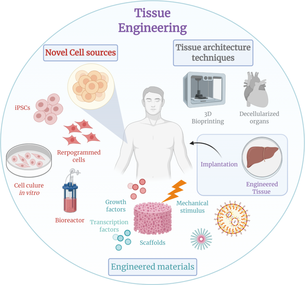
Bioreactors
Bioreactor based 3D models constitute another innovative platform that aids in answering fundamental research questions. Bioreactor-based 3D cell culture models are being optimized in combination with computational modeling and novel sensor technologies to improve basic physiology and tissue development research with the hope of serving as a solid platform to streamline significant bioprocesses needed for safe and aptly scaled tissue production of translational research (3,7).
Organ-on-a-chip (OoC)
The term organ on-a-chip refers to microfluidic devices for the cultivation of living cells. These devices can be applied to investigate organ development and mimic in vivo-like metabolic and physiological cell behavior. OoCs are particularly useful in studying the crosstalk between organs. The reproducible and modifiable nature of OoCs make them promising tools for the future use in drug discovery and development research, further reducing the need for costly animal models (4).,7
Bioprinting
Bioprinting is a novel technique that was introduced and supported by bioengineering technologies. to the term bioprinting refers to a technique that uses cells as well as other material for scaffold production as a type of “bioink” to print a biological cell or organ. The potential of bioprinting in regenerative medicine is still under investigation (5).
References
1. Duval K., Grover H., Han L.-H., Mou Y., Pegoraro A. F., Fredberg J., et al. (2017). Modeling Physiological Events in 2D vs. 3D Cell Culture. Physiology 32 (4), 266–277.
2. Drost J., Clevers H. (2018). Organoids in Cancer Research. Nat. Rev. Cancer 18 (7), 407–418.
3. Wendt D., Riboldi S. A., Cioffi M., Martin I. (2009). Potential and Bottlenecks of Bioreactors in 3D Cell Culture and Tissue Manufacturing. Adv. Mat. 21 (32–33), 3352–3367.
4. Bhatia S. N., Ingber D. E. (2014). Microfluidic Organs-On-Chips. Nat. Biotechnol. 32 (8), 760–772.
5. Arslan-Yildiz A., Assal R. E., Chen P., Guven S., Inci F., Demirci U. (2016). Towards Artificial Tissue Models: Past, Present, and Future of 3D Bioprinting. Biofabrication 8 (1), 014103.
6. Matai I., Kaur G., Seyedsalehi A., McClinton A., Laurencin C. T. (2020). Progress in 3D Bioprinting Technology for Tissue/organ Regenerative Engineering. Biomaterials 226, 119536.
7. Cacciamali A, Villa R and Dotti S (2022) 3D Cell Cultures: Evolution of an Ancient Tool for New Applications. Front. Physiol. 13:836480.