Epithalial cells for spheroid formation
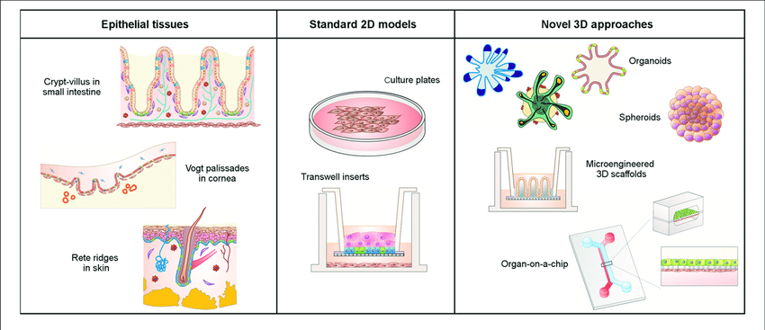
The Epithelium is a thin, continuous, layer of tightly packed cells formed as complex 3D structures such as cysts, tubules or invaginations. The shape of the epithelium is essential for its function as it aids in creating biochemical gradients that directs cell placement and compartmentalization. In addition to providing protection to underlying tissues, all epithelial cells perform a wide array of vital functions such as diffusion, filtration, secretion, trans cellular transport etc. that is crucial for proper organ function. The adult stem cells present within epithelial tissues ensure rapid repair and regeneration of cells (Crosnier et al, 2006, Torras et al, 2018, Vrana et al, 2013) .
Epithelial cell models used in Patho-physiological research
There is a wide range of systems currently utilized for basic function studies, disease modeling, drug discovery, and tissue regeneration studies; albeit some being more effective than others. There are conventional animal models that are used to study basic, in-vivo physiology and tissue interactions, but are unable to provide translatable information on human response due to species differences. 2D cell culture models are able to simulate early cellular response, but have poor predictive capabilities (Griffith et al, 2006). 3D culture technology has helped to overcome these limitations, as it provides native state structures on a reliable, reproducible in-vitro system. There are many application-specific 3D models of epithelial tissues, ranging from self-assembled cultures (i.e. spheroids or organoids), lab on chip devices, and engineered micro tissues (Torras et al, 2018).
Self-assembled 3D cultures
Spheroids and organoids are now widely used as reliable in vitro tools for drug testing and disease modeling. Organoids are especially valuable in basic science and patient-specific disease research. They provide complex 3D cell culture systems that better reflect native tissue composition, structure, and function.
Spheroids are also important in cancer research. Their spherical structure helps reproduce the biochemical and cellular environment found in many tumors (Lin et al., 2008). For this reason, researchers often generate tumor spheroids from primary cancer cells, including glioma, breast, colon, ovarian, and prostate cancer cells (Ishiguro et al., 2017).
Several breast cancer cell lines can be cultured as spheroids, including D492HER, D492, MCF10A, MDA-MB231, MCF-7, and HCC143. In addition, epidermoid carcinoma cell lines such as A431 can also form spheroid cultures. These models offer a reliable and adaptable system for pharmacological studies.
Moreover, spheroid cultures are relatively simple to establish and cost-effective to maintain. They are also reproducible and easy to modify. As a result, they are well suited for high-throughput drug screening.
References
- Vrana, N. E., Lavalle, P., Dokmeci, M. R., Dehghani, F., Ghaemmaghami, A. M., and Khademhosseini, A. (2013). Engineering functional epithelium for regenerative medicine and in vitroorgan models: a review. Tissue Eng. Part B Rev. 19, 529–543. doi: 10.1089/ten.teb.2012.0603
- Crosnier, C., Stamataki, D., and Lewis, J. (2006). Organizing cell renewal in the intestine: stem cells, signals and combinatorial control. Rev. Genet.7, 349–359. doi: 10.1038/nrg1840
- Lin, R. Z., and Chang, H. Y. (2008). Recent advances in three-dimensional multicellular spheroid culture for biomedical research. J.3, 1172–1184. doi: 10.1002/biot.200700228
- Ishiguro, T., Ohata, H., Sato, A., Yamawaki, K., Enomoto, T., and Okamoto, K. (2017). Tumor-derived spheroids: relevance to cancer stem cells and clinical applications. Cancer Sci.108, 283–289. doi: 10.1111/cas.13155
- Griffith, L. G., and Swartz, M. A. (2006). Capturing complex 3D tissue physiology in vitro. Rev. Mol. Cell Biol.7, 211–224. doi: 10.1038/nrm1858