Bioprinting for cardiac tissue engineering
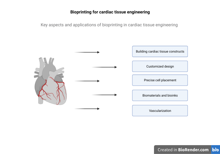
Bioprinting is an innovative technology with immense potential in cardiac tissue engineering. It offers precise control over cell placement and biomaterial deposition, making it a promising approach for creating functional cardiac tissue constructs. Here are some key aspects and applications of bioprinting in cardiac tissue engineering: Building cardiac tissue constructs Bioprinting allows researchers to create 3D cardiac tissue constructs layer by layer. These constructs can mimic the structural and functional characteristics of native heart tissue, including cardiomyocytes, blood vessels, and extracellular matrix components.
Customized design
Bioprinting enables the customization of cardiac tissue constructs based on a patient’s specific needs. This personalized approach is essential for addressing individual variations in cardiac anatomy and pathology.
Precise cell placement
Bioprinters can position cardiomyocytes and other cell types with high precision. This precision is crucial for creating organized cardiac structures, including muscle fibers and blood vessels (3).
Biomaterials and bioinks
Specially formulated bioinks, often containing hydrogels, are used to encapsulate and support cardiac cells during bioprinting. These bioinks provide a suitable microenvironment for cell growth and tissue development.
Vascularization
Bioprinting techniques are employed to create intricate vascular networks within cardiac tissue constructs. This is critical for ensuring proper nutrient and oxygen supply to the engineered tissue.
Electrical
integration The electrical integration of bioprinted cardiac tissues is a key focus. Bioprinting can be used to position cells and materials with specific electrical properties to promote synchronous beating and electrical signaling, mimicking the heart’s natural behavior.
While bioprinting for cardiac tissue engineering is a promising field, there are challenges to address, including the need for vascularization and long-term functionality. Ongoing research and technological advancements continue to drive progress in this exciting area, with the ultimate goal of providing effective treatments for heart disease and improving patient outcomes.
References
1. Wang Z, Wang L, Li T, Liu S, Guo B, Huang W, Wu Y. 3D bioprinting in cardiac tissue engineering. Theranostics. 2021 Jul 6;11(16):7948-7969. doi: 10.7150/thno.61621. PMID: 34335973; PMCID: PMC8315053.
2. Wu, Catherine A., Yuanjia Zhu, and Y. Joseph Woo. 2023. “Advances in 3D Bioprinting: Techniques, Applications, and Future Directions for Cardiac Tissue Engineering” Bioengineering 10, no. 7: 842. https://doi.org/10.3390/bioengineering10070842
3. Sung K, Patel NR, Ashammakhi N, Nguyen KL. 3-Dimensional Bioprinting of Cardiovascular Tissues: Emerging Technology. JACC Basic Transl Sci. 2021 May 24;6(5):467-482. doi: 10.1016/j.jacbts.2020.12.006. PMID: 34095635; PMCID: PMC8165127.
4. Akter F, Araf Y, Naser IB, Promon SK. Prospect of 3D bioprinting over cardiac cell therapy and conventional tissue engineering in the treatment of COVID-19 patients with myocardial injury. Regen Ther. 2021 Dec;18:447-456. doi: 10.1016/j.reth.2021.09.007. Epub 2021 Sep 30. PMID: 34608441; PMCID: PMC8481096.
5. Kato B, Wisser G, Agrawal DK, Wood T, Thankam FG. 3D bioprinting of cardiac tissue: current challenges and perspectives. J Mater Sci Mater Med. 2021 May 6;32(5):54. doi: 10.1007/s10856-021-06520-y. PMID: 33956236; PMCID: PMC8102287