Different approaches for 3D primary cell culture model development
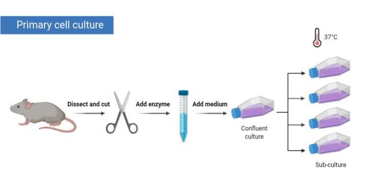
Primary cell cultures are ex-vivo cultures produced from cells that are freshly harvested from a multicellular organism. Generally, primary cell cultures are more representative of in-vivo tissues than immortalized cell lines (1). However culturing primary cells are not without its difficulties. They require adequate media and nutrient conditions to thrive and after number of cell divisions, the cells adopt a senescent phenotype, resulting in an irreversible cell cycle arrest (2). Immortalized cell lines are thus generated to over come these limitations.
Primary cells can become immortalized spontaneously as observed in the case of HeLa cells or through genetic manipulation such as in HEK cells. Once immortalized, the viability of cells is extended as they can then be subcultured indefinitely (3).
Advantages of Primary cell cultures
Primary cell cultures gained more popularity in the 2000s, due to the several advantages it had over immortalized cell lines (4-6).
- Mimicked in-vivo like cellular heterogeneity, with a transcriptional and proteomic profile consistent with native state, especially when cultured in a 3D platform.
- The in-vivo like cellular structure and function lead to more realistic functional response to external variables and drug responses.
- Avoids cellular homogeneity through natural selections, and genetic aberrations that are typically observed in immortalized cell lines.
2D and 3D primary cell cultures
2D cell cultures were the go-to method to study cells for the past century. While it has been a vital tool in advancing the knowledge of cellular behavior, it consisted of many limitations that have reduced its use in research. 2D cultures are unable to accurately capture the cellular structure and function found in in-vivo conditions. 3D cultures on the other hand has stepped into fill the gap, and provided a modifiable, and reliable platform to generate cells that mimic cellular structure and function present in-vivo. 3D cultures have thus advanced the field of cancer and stem cell research, drug discovery and disease modeling (7).
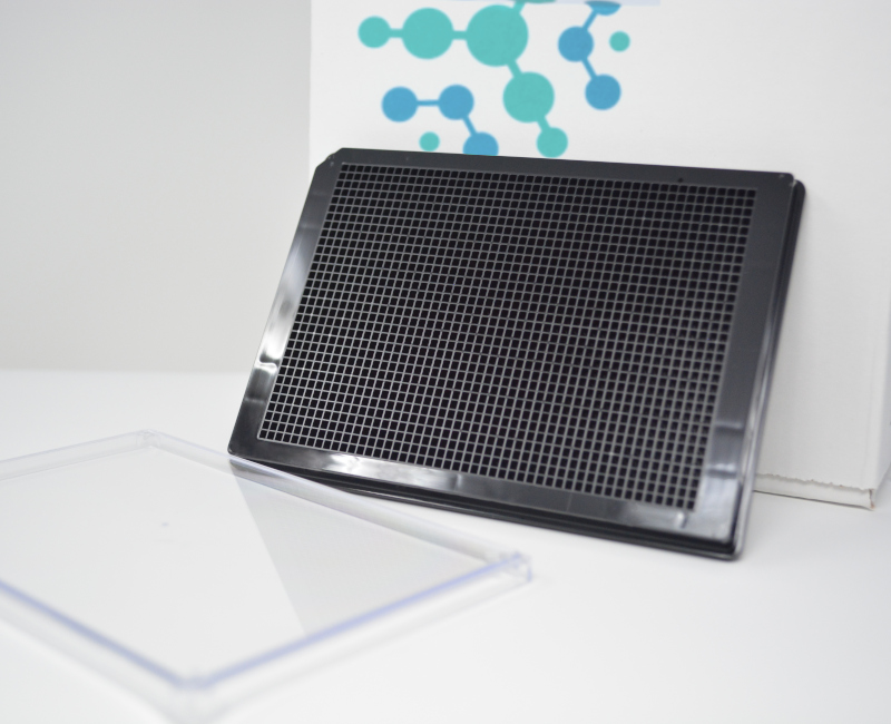
In 3D cell cultures cells are cultured with a supporting scaffold (such as hydrogel), or without a scaffold with the use of hanging drop method, magnetic levitations or spheroid microplates with specialized cell repellant polymeric coating (such as BioFloatTM). Primary 3D cultures have been particularly successful in both types of 3D cultures. With the inherent ability of primary cells to mimic natural cellular behavior, 3D culturing techniques have aided in creating an in-vivo like cell state with intact transcriptional and proteomic profiles, thus providing an excellent platform for tumor research, stem cell research, neural disease modeling and drug screens (7).
References
1. Geraghty, R J; Capes-Davis, A; Davis, J M; Downward, J; Freshney, R I; Knezevic, I; et al. (September 2014). “Guidelines for the use of cell lines in biomedical research”. British Journal of Cancer. 111 (6): 1021–1046.
2. Campisi, Judith; d’Adda di Fagagna, Fabrizio (September 2007). “Cellular senescence: when bad things happen to good cells”. Nature Reviews Molecular Cell Biology. 8 (9): 729–740.
3. Freshney, R. Ian; Freshney, Mary G., eds. (1996). Culture of immortalized cells. New York: Wiley-Liss. ISBN 978-0-471-12134-3.
4. Gillet, J.-P.; Varma, S.; Gottesman, M. M. (3 April 2013). “The Clinical Relevance of Cancer Cell Lines”. JNCI Journal of the National Cancer Institute. 105 (7): 452–458. doi:10.1093/jnci/djt007. PMC 3691946. PMID 23434901.
5. Cree, Ian A; Glaysher, Sharon; Harvey, Alan L (August 2010). “Efficacy of anti-cancer agents in cell lines versus human primary tumour tissue”. Current Opinion in Pharmacology. 10 (4): 375–379. doi:10.1016/j.coph.2010.05.001. PMID 20570561.
6. Tiriac, Hervé; Belleau, Pascal; Engle, Dannielle D.; Plenker, Dennis; Deschênes, Astrid; Somerville, Tim D. D.; et al. (September 2018). “Organoid Profiling Identifies Common Responders to Chemotherapy in Pancreatic Cancer”. Cancer Discovery. 8 (9): 1112–1129.
7. Jensen C and Teng Y (2020) Is It Time to Start Transitioning From 2D to 3D Cell Culture? Front. Mol. Biosci. 7:33
8. Gaebler, Manuela & Silvestri, Alessandra & Haybaeck, Johannes & Reichardt, Peter & Lowery, Caitlin & Stancato, Louis & Zybarth, Gabriele & Regenbrecht, Christian. (2017). Three-Dimensional Patient-Derived In Vitro Sarcoma Models: Promising Tools for Improving Clinical Tumor Management. Frontiers in Oncology. 7. 10.3389/fonc.2017.00203.


