Spheroids and Organoids on organ-on-chip platform
In the recent decades, 3D bioprinting methods have been widely used in tissue engineering and regenerative medicine. Additionally, these techniques have also emerged as a novel approach in the development of in vivo-like 3D cell culture models (1-3).
Advantages of 3D bioprinting techniques
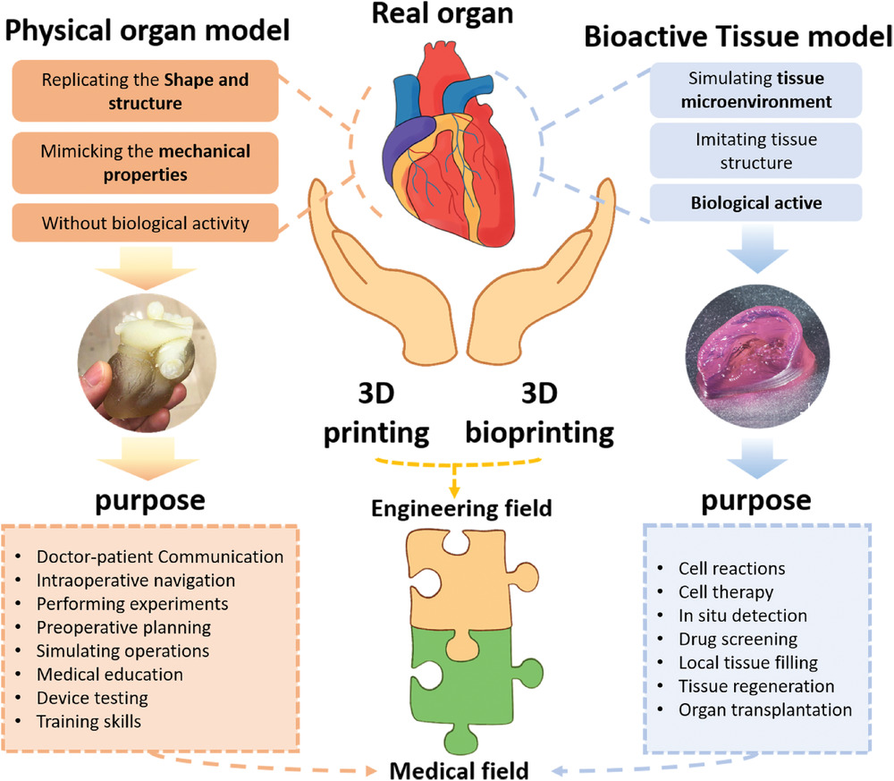
Three dimensional (3D) bioprinting is the use of 3D printing–like techniques to combine cells, growth factors and other biomaterials in order to produce 3D models with in vivo-like physiological properties.
3D bioprinting techniques are compatible with high throughput screening of bio active models, are able to create chemical and physical gradients, and allows for co-culture of cells through the introduction of biomaterial-encased living cells (bioink) with custom made structures. Such advantageous aspects of 3D bioprinting help to overcome some of the limitations associated with organoids (miniature self-assembled 3D structures mimicking the structure and function of human organs), such as their limited size and reproducibility (3,4).
3D bioprinting techniques have been explored in diverse ways to optimize the efficiency and accuracy of available 3D culture models. Daly and colleagues incorporated 3D printed spheroids into self-healing support hydrogels yielding microtissues that can subsequently be used for in vitro experiments. Others studies report spheroids formation inside porous composite granular hydrogels and their positioning in a 3D printer, to produce effectively reorganized 3D constructs (5,6).
However, despite their high reproducibility, these techniques also have significant limitations. These challenges include the incorporation of vasculature in organ models, as well as in obtaining the right material to produce accurate constructs of co-cultured cells. In tumor research models, organoids maintained at static conditions perform poorly over a prolonged period of time, as oxygen and nutrient exchange becomes insufficient. Moreover, tumor cells don’t encounter shear stress which in turn affects their ability to metastasize (3).
Combining Microfluidics with organ-on-chip
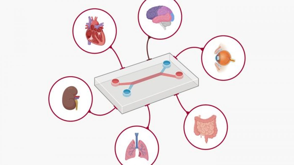
As an attempt to overcome the limitations of bioprinting methods, researchers combined microfluidic technologies with cell culture leading to the production of an advanced preclinical platform called organ-on-chip (OoC). OoC offer integration of cell culture models, enable more effective nutrient exchange, and allow incorporation of sensors, actuators, and automation systems. Furthermore, these devices are also able to create many of the physical and chemical stimuli essential for recreating in vivo-like physiology thus yielding translatable data needed for pre-clinical evaluations.
Compared to animal models, Organ-on Chip methods are also relatively low cost and show high reproducibility rates thus reducing the overall cost, labor, and time (7-10). The first organ-on-chip model was produced in 2010 and consisted on a Lung-on-a-Chip. Since then, a wide range of OoCs have been developed such as liver, lung, kidney, heart, breast tissue as well as combination of several organs. Collectively organ-on-chip is proving to be breakthrough in the field of developing 3D models for biomedical research (3,11).
References
1. N.S. Bhise, V. Manoharan, S. Massa, A. Tamayol, M. Ghaderi, A liver-on-a-chip platform with bioprinted hepatic spheroids, Biofabrication 8, 2016.
2. R.O. Rodrigues, P.C. Sousa, J. Gaspar, M. Ban ̃obre-Lo ́pez, R. Lima, G. Minas, Organ-on-a-Chip: a preclinical microfluidic platform for the progress of nanomedicine, Small 16, 2020.
3. Violeta Carvalho, Manuel Bañobre-López, Graça Minas, Senhorinha F.C.F. Teixeira, Rui Lima, Raquel O. Rodrigues. The integration of spheroids and organoids into organ-on-a-chip platforms for tumour research: A review. Bioprinting, Volume 27, 2022.
4. R. Burdis, D.J. Kelly, Biofabrication and bioprinting using cellular aggregates, microtissues and organoids for the engineering of musculoskeletal tissues, Acta Biomater. 126, 2021,1–14.
5. C. Norotte, F.S. Marga, L.E. Niklason, G. Forgacs, Scaffold-free vascular tissue engineering using bioprinting, Biomaterials 30, 2009, 5910–5917.
6. A.C. Daly, M.D. Davidson, J.A. Burdick, 3D bioprinting of high cell-density heterogeneous tissue models through spheroid fusion within self-healing hydrogels, Nat. Commun. 12, 2021, 1–13.
7. Q. Wu, J. Liu, X. Wang, L. Feng, J. Wu, X. Zhu, W. Wen, X. Gong, Organ-on-a- chip: recent breakthroughs and future prospects, Biomed. Eng. Online 19, 2020, 1–19.
8. R. Kalot, R. Mhanna, R. Talhouk, Organ-on-a-chip platforms as novel advancements for studying heterogeneity, metastasis, and drug efficacy in breast cancer, Pharmacol. Ther. 237 2022, 108156.
9. G.M. Whitesides, The origins and the future of microfluidics, Nature 442, 2006, 368–373.
10. D.Y. Park, J. Lee, J.J. Chung, Y. Jung, S.H. Kim, Integrating organs-on-chips: multiplexing, scaling, vascularization, and innervation, Trends Biotechnol. 38, 2020, 99–112.
11. D. Huh, B.D. Matthews, A. Mammoto, M. Montoya-Zavala, H. Yuan Hsin, D. E. Ingber, Reconstituting organ-level lung functions on a chip, Science 328, 2010, 1662–1668.