Organoid Models Of Genetic Diseases
Organoid models are a popular and reliable method in 3D cell culture. Organoids are 3D cellular structures, derived from either stem cells or organ specific progenitor cells. When provided with an external scaffold like Matrigel, they form a complex cluster of cells that later differentiate to form a miniature version of an organ, able to mimic in vivo-like cellular architecture and function (1,2). Such organoid models are now increasingly being used to model genetic diseases.
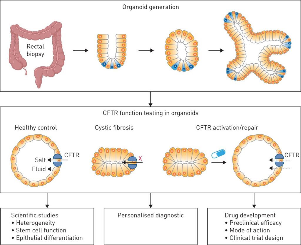
Intestinal organoids for Cystic Fibrosis
In the recent years two main types of organoids have been generated to model genetic diseases. One type of organoids is generated using cells derived from patient biopsies; the other type is represented by genetically engineered wild type organoids. These two methods have been primarily employed to study the effect of lethal mutations during early life development.
In 2013, Dekkers and colleagues were able to develop a test investigating the effect of Forskolin treatment on healthy intestinal organoids and those generated from rectal biopsies of cystic fibrosis (CF) patients. The healthy organoids showed rapid swelling in response to Forskolin, whereas a reduced response was shown in CF organoids . This test has proved very reliable to investigate drug response in the context of cystic fribrosis and has become the first personalized organoid medicine application for cystic fibrosis in the Netherlands (3).
Hepatic organoid models
Other types of organoids obtained with cells derived from patient suffering from genetic diseases include hepatic organoids from 1-antitrypsin deficient patients as well as from patients with Alagille syndrome. Both proved successful in representing the respective genetic defects (4,5). These studies successfully proved the reliability and accuracy of organoids in replicating disease characteristics in an in vitro setting and thus are an indispensable resource for basic physiological studies, and patient specific therapy development.
References
1. Clevers H. (2016). Modeling Development and Disease with Organoids. Cell. 165 (7), 1586–1597.
2. Cacciamali A, Villa R and Dotti S (2022) 3D Cell Cultures: Evolution of an Ancient Tool for New Applications. Front. Physiol. 13:836480.
3. Dekkers, J. F., Wiegerinck, C. L., De Jonge, H. R., Bronsveld, I., Janssens, H. M., De Winter-de Groot, K. M., et al. (2013). A Functional CFTR Assay Using Primary Cystic Fibrosis Intestinal Organoids. Nat. Med. 19 (7), 939–945.
4. Huch, M., Gehart, H., Van Boxtel, R., Hamer, K., Blokzijl, F., Verstegen, M. M., et al. (2015a). Long-term Culture of Genome-Stable Bipotent Stem Cells from Adult Human Liver. Cell. 160 (1–2), 299–312.
5. Andersson, E. R., Chivukula, I. V., Hankeova, S., Sjöqvist, M., Tsoi, Y. L., Ramsköld, D., et al. (2018). Mouse Model of Alagille Syndrome and Mechanisms of Jagged1 Missense Mutations. Gastroenterology 154 (4), 1080–1095.
6. Van Mourik P, Beekman JM, van der Ent CK. Intestinal organoids to model cystic fibrosis. Eur Respir J 2019; 54: 1802379.