Necrosis in spheroids and how to detect it
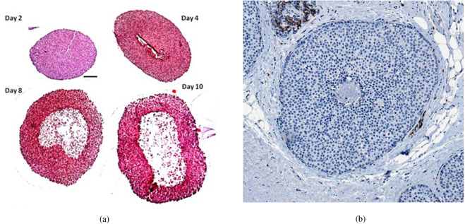
As spheroids grow, oxygen diffusion from the external layers of cells into the core gets limited and this leads to cellular stress, energy deprivation, and eventually necrosis. The development of necrosis in spheroids depends on various factors, including the spheroid size, cell type, culture conditions, and the absence of an appropriate vascular network.
Choosing advanced culture systems like BIOFLOATTM plates to generate round and uniform spheroids, incorporating supportive cells such as endothelial cells to establish a vascular-like network, and employing different culture media and growth factors promote spheroid viability and help in mitigating necrosis development.
Necrosis can be detected through various methods. This article gives an overview of five commonly used techniques:
-
- Visual observation: Necrotic regions within a spheroid may appear darker or discolored compared to the surrounding healthy tissue. Signs of necrosis such as changes in cell morphology or loss of cellular structure can be observed under a microscope (1).
- Viability assays: Viability assays are used to measure cell metabolic activity or cell membrane integrity. For example, the MTT (3-(4,5-Dimethylthiazol-2-yl)-2,5-diphenyltetrazolium bromide) assay or the resazurin assay can be used. These assays rely on the conversion of a colorless substrate into a colored or fluorescent product by viable cells. Necrotic cells show reduced metabolic activity and thus lower signal intensity (2).
- Fluorescent staining: Fluorescent dyes, such as propidium iodide (PI), can be used to identify necrotic areas within a spheroid. PI is impermeable to live cells but stains the nuclei of dead or dying cells. Spheroids are incubated with PI and then visualized under the fluorescence microscope. Necrotic regions will appear bright due to the stained nuclei.
- Lactate Dehydrogenase (LDH) assay: Lactate dehydrogenase, or LDH, is a stable and soluble cytosolic enzyme present in most eukaryotic cells. LDH is released into the culture medium when cells undergo necrosis. Measuring the levels of LDH in the culture medium surrounding the spheroid helps in identifying the severity of necrosis (3).
- Immunohistochemistry: Specific antibodies can be used to target markers associated with necrosis, such as cleaved caspase-3 or high mobility group box 1 protein (HMGB1). By staining the spheroid with these antibodies and visualizing the results, necrotic regions can be identified. Additionally, miR-122 has been shown to be a highly sensitive marker for necrotic cell death (4).
Combining multiple techniques and validating the results with other methods or conducting further analysis, such as histological examination, can provide a more comprehensive evaluation of necrosis within a spheroid.
References:
- Tinari A, Giammarioli AM, Manganelli V, Ciarlo L, Malorni W. Analyzing morphological and ultrastructural features in cell death. Methods Enzymol. 2008;442:1-26. doi: 10.1016/S0076-6879(08)01401-8. PMID: 18662562.
- Kumar P, Nagarajan A, Uchil PD. Analysis of Cell Viability by the MTT Assay. Cold Spring Harb Protoc. 2018 Jun 1;2018(6). doi: 10.1101/pdb.prot095505. PMID: 29858338.
- Chan FK, Moriwaki K, De Rosa MJ. Detection of necrosis by release of lactate dehydrogenase activity. Methods Mol Biol. 2013;979:65-70. doi: 10.1007/978-1-62703-290-2_7. PMID: 23397389; PMCID: PMC3763497.
- Bogajewska-Rylko E, Ahmadi N, Pyrek M, Dziekan B, Filas V, MarszaŁek A, Szylberg L. When Is Immunohistochemistry Useful in Assessing Tumor Necrotic Tissue? Anticancer Res. 2021 Jan;41(1):197-201. doi: 10.21873/anticanres.14765. PMID: 33419813.


