Miniature Spheroid Formation In 384 Well Plates
3D cultures have become a vital tool in tumor related research and drug discovery. 3D cultures of tumor cell lines bear remarkable structural and physiological similarities to in vivo solid tumors. Spheroids of cancer cells are able to mimic the complex in-vivo structures of tumors, cell-cell interactions, zones of proliferating and quiescent cells, and oxygen and nutrient gradients, compared to traditional 2D cultures. Thus spheroids are considered a relevant model for tumor pathophysiological studies as well as drug efficacy and toxicity testing (1).
Spheroid cultures for high throughput screening (HTS)
Most standardized spheroid protocols have been optimized for use in high throughput/high content screening. However, much improvement is still being made to reduce well-well variability, in spheroid size and shape. Liquid overlay, agarose coated plates, and low attachment plates are common ways of spheroid culture techniques (4). These methods are low cost, and low tech, and easily adaptable from standard multi-well plates to micro-titer plates. Spheroid formation in 96 well plates, and 384 well plates has proven effective at being used in HTS (4,5). Albeit edge effect is a common challenge, it is easily overcome with improved culture conditions that prevent the loss of media, and reduce the well-well variability (1-3,6-8).
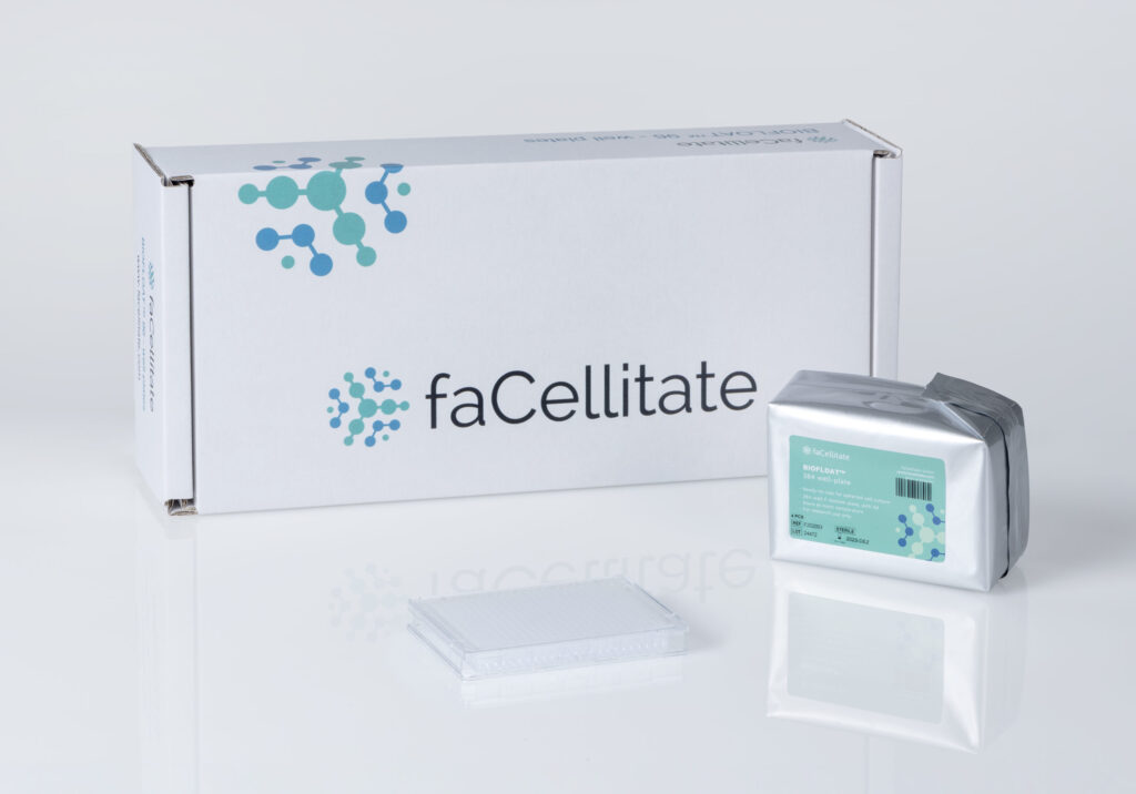
Spheroid formation in low attachment 384 well plates
To advance the methods of forming scaffold free, round miniature spheroids, Facellitate™ launched its own low attachment 384 well plates. These plates are coated with specialized polymer coating that renders the plate surface cell repellant, encouraging the self-aggregation of cells, leading to the formation of round spheroids in a relatively short period of time. Mouse fibroblasts 3T3 cells, seeded at 3000 cells/well led to the formation of single round spheroids in over 95% of the wells. The relatively short time required for spheroid formation, and the low variability in spheroid shape and size across wells make these plates an excellent low, cost, low tech option that can be easily adapted on a high throughput platform for drug response and toxicity screens (9).
References
1. Das, V.; Bruzzese, F.; Konečný, P.; et al. Pathophysiologically Relevant In Vitro Tumor Models for Drug Screening. Drug Discov. Today 2015, 20, 848–855.
2. LaBarbera, D. V.; Reid, B. G.; Yoo, B. H. The Multicellular Tumor Spheroid Model for High-Throughput Cancer Drug Discovery. Expert Opin. Drug Discov. 2012, 7, 819–830.
3. Monjaret, F.; Fernandes, M.; Duchemin-Pelletier; et al. Fully Automated One-Step Production of Functional 3D Tumor Spheroids for High-Content Screening. J. Lab. Autom. 2016, 21, 268–280
4. Wenzel, C.; Riefke, B.; Gründemann, S.; et al. 3D HighContent Screening for the Identification of Compounds That Target Cells in Dormant Tumor Spheroid Regions. Exp. Cell Res. 2014, 323, 131–143.
5. Li, Q.; Chen, C.; Kapadia, A.; et al. 3D Models of EpithelialMesenchymal Transition in Breast Cancer Metastasis: HighThroughput Screening Assay Development, Validation, and Pilot Screen. J. Biomol. Screen. 2011, 16, 141–154.
6. Berg, M.; Undisz, K.; Thiericke, R.; et al. Evaluation of Liquid Handling Conditions in Microplates. J. Biomol. Screen. 2001, 6, 47–56
7. Berthier, E.; Warrick, J.; Yu, H.; et al. Managing Evaporation for More Robust Microscale Assays Part 1. Volume Loss in High Throughput Assays. Lab. Chip 2008, 8, 852–859.
8. Walzl, A.; Kramer, N.; Mazza, G.; et al. A Simple and Cost Efficient Method to Avoid Unequal Evaporation in Cellular Screening Assays, Which Restores Cellular Metabolic Activity. Int. J. Appl. Sci. Technol. 2012, 2, 17–25. 9. https://facellitate.com/product/biofloat-384-well-plates/