Bone microenvironment
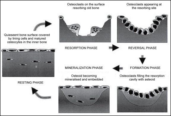
Bone is a mineralized connective tissue composed of four types of cells namely, osteoblasts, osteocytes, osteoclasts and bone lining cells. Bone tissue is constantly undergoing remodeling via the bone resorption by osteoclasts and bone formation by osteoblasts. Osteocytes operate as the mechanosensors and guide the bone re modeling process (Rinaldo et al, 2015). This delicately balanced process is carefully regulated by local and systemic factors. An imbalance between resorption and formation can result in bone diseases such as osteoporosis (Bicer et al, 2021). Cell lines derived from bone are typically cultured in the lab in 2D monolayer form, albeit with limitations.
Mimicking bone microenvironment in the lab
Replicating the bone microenvironment is essential to obtain data that is physiologically relevant and translatable. Recent studies have thus shifted its focus from using traditional 2D cell culture models to novel and complex 3D models that include, spheroids, cell sheets, hydrogels, bioreactors, and microfluidic chips (Mohammed AM, 2008).
3D spheroids
Spheroids are simple self-assembled cell aggregate 3D cell culture system with no supporting material needed for cell growth. The spheroid culture system conserves cell viability, and facilitates the intracellular and cell-matrix networks, creating a in-vivo like microenvironment, with signaling molecules such as cytokines, chemokines and angiogenic factors that is present in native physiological state (Mironov et al, 2009). Furthermore 3D spheroids can also be co-cultured with other cell lines in different densities to better replicate the in-vivo conditions. For e.g. osteoblasts can be co cultured with human umbilical vascular endothelium cells (huVECs) in 96 well-plates with specialized coating to form uniform spheroids with separated cell populations within a single spheroid . Therefore 3D cell culture models such as the spheroid systems are an excellent platform that is rapid, reliable and reproducible and can be easily modified by changing different parameters (i.e. initial seeding density, cell line) to generate cells that are biologically relevant and able to yield clinically significant, translatable data for disease or pharmaceutical research.
References
- Rinaldo Florencio-Silva, Gisela Rodrigues da Silva Sasso, Estela Sasso-Cerri, Manuel Jesus Simões, Paulo Sérgio Cerri, “Biology of Bone Tissue: Structure, Function, and Factors That Influence Bone Cells”, BioMed Research International, vol. 2015, Article ID 421746, 17 pages, 2015. https://doi.org/10.1155/2015/421746
- Bicer, M., Cottrell, G.S. & Widera, D. Impact of 3D cell culture on bone regeneration potential of mesenchymal stromal cells. Stem Cell Res Ther12, 31 (2021). https://doi.org/10.1186/s13287-020-02094-8
- Mohamed AM. An overview of bone cells and their regulating factors of differentiation. Malays J Med Sci. 2008;15(1):4-12.
- Mironov, V., Visconti, R. P., Kasyanov, V., Forgacs, G., Drake, C. J., and Markwald, R. R. (2009). Organ Printing: Tissue Spheroids as Building Blocks. Biomaterials30 (12), 2164–2174. doi:10.1016/j.biomaterials.2008.12.084
- https://facellitate.com/wp-content/uploads/Establishment-of-perfect-3D-spheroids-for-1.pdf


