The Endothelium: An interface between Blood, tissue and lymph
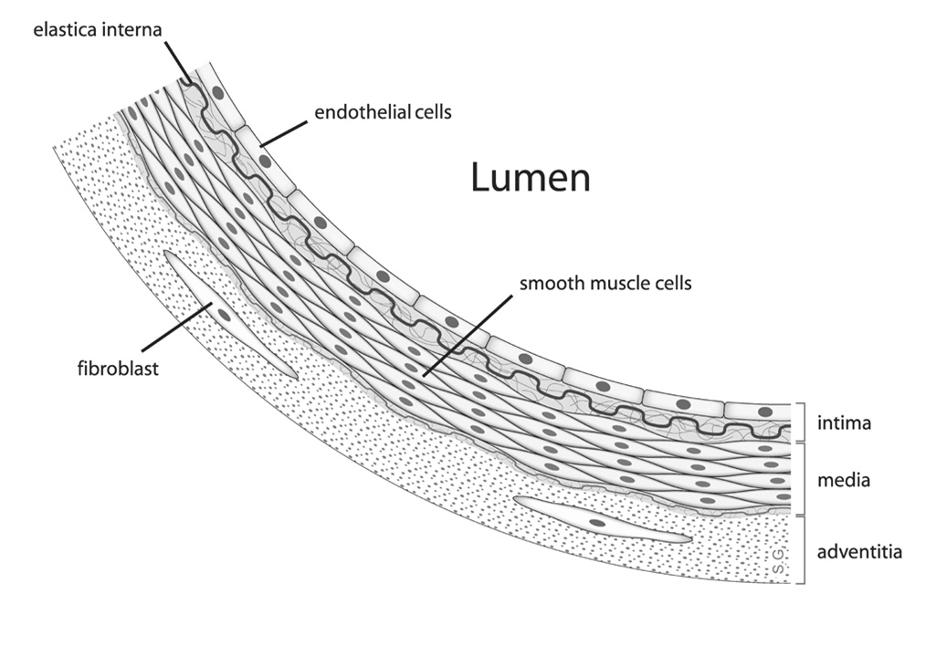
The Endothelium consists of a single layer of squamous endothelial cells, forming the inner lining of the circulatory system. It is has varying degrees of permeability depending on the organ physiology as it operates as a barrier between vessels and tissues. Thus, the endothelial cells play an important role in fluid transport between vessels and tissues, angiogenesis, tissue repair, hormone trafficking and inflammatory response (Michiels C, 2003). The vascularization, and high nutrient supply within, also makes the endothelium an ideal site for tumors. In the lab, these cells can easily cultured in a 2D monolayer form, or in a 3D culture system (spheroid or organoids) for a comprehensive cellular structure that mimics the native state.
Pathology
Impaired endothelial function is considered a strong indicator of vascular disease, such as atherosclerosis, hypertension and thrombosis (He et al, 2017., Gimbrone and García-Cardeña, 2016). In addition to coronary disease, endothelial dysfunction presents as an early sign of autoimmune diseases such as rheumatoid arthritis and Lupus. While the endothelial lining provides protection, it is vulnerable to some incoming pathogens such as Kaposi’s sarcoma-associated herpes virus (KSHV), often linked to tumors such as Kaposi’s sarcoma and primary effusion lymphoma (Ojala, et al, 2014).
Endothelial cells are anchored to the tissues via the basement membrane; a thin sheet of the extracellular matrix. Typically when the cells are removed for monolayer cultures, they lose contact with the basement membrane, consequently resulting in cell behaviors that are different to that in its native environment, yielding untranslatable data for pharmacological studies. Therefore much effort has gone in to optimizing the 3D culture systems of endothelial cells in physiological and pathological contexts (Heiss et al, 2015).
Spheroids of endothelial cells
Spheroids consist of self-assembled aggregates of cells that are able to develop key components necessary for physiologically relevant spatial growth as well as cell-cell interactions making it an excellent choice as a 3D model system. To this end, human umbilical vein endothelial cells (HuVECs) are used in spheroid cultures in studying endothelial cell function, angiogenesis and pathogen interactions (Finkenzeller et al, 2009., Heiss et al, 2015).
Immortalized endothelial cells originating from HUVEC cells (HuARLT cells) are commonly used to study tumor cell states in Kaposi’s sarcoma (Lipps et al, 2017). Both these cell types can be effectively cultured as a spheroid in ultra low attachments U-bottom plates with specialized polymer coating providing a low cost, highly reproducible system applicable for physiological studies as well as for high throughput assays (https://facellitate.com/wp-content/uploads/FAQs_new-2.pdf).
References
1. Michiels C (2003) Endothelial cell functions. J Cell Physiol 196(3):430–443. doi:10.1002/jcp.10333
2. He L, Huang X, Kanisicak O, et al. Preexisting endothelial cells mediate cardiac neovascularization after injury. J Clin Invest. 2017;127(8):2968-2981. doi:10.1172/JCI93868
3. Gimbrone MA Jr, García-Cardeña G. Endothelial Cell Dysfunction and the Pathobiology of Atherosclerosis. Circ Res. 2016;118(4):620-636. doi:10.1161/CIRCRESAHA.115.306301
4. Ojala PM, Schulz TF (2014) Manipulation of endothelial cells by KSHV: implications for angiogenesis and aberrant vascular differentiation. Semin Cancer Biol. doi:10.1016/j.semcancer. 2014.01.008
5. Finkenzeller, G., Graner, S., Kirkpatrick, C. J., Fuchs, S., & Stark, G. B. (2009). Impaired in vivo vasculogenic potential of endothelial progenitor cells in comparison to human umbilical vein endothelial cells in a spheroid-based implantation model. Cell proliferation, 42(4), 498–505. https://doi.org/10.1111/j.1365-2184.2009.00610.x
6. Heiss M, Hellström M, Kalén M, May T, Weber H, Hecker M, Augustin HG, Korff T. Endothelial cell spheroids as a versatile tool to study angiogenesis in vitro. FASEB J. 2015 Jul;29(7):3076-84. doi: 10.1096/fj.14-267633. Epub 2015 Apr 9. PMID: 25857554.
7. Lipps, C., Badar, M., Butueva, M. et al. Proliferation status defines functional properties of endothelial cells. Cell. Mol. Life Sci. 74, 1319–1333 (2017). https://doi.org/10.1007/s00018-016-2417-5
8. TriggleChris R., Samuel Samson Mathews, Ravishankar Shalini, MareiIsra, Arunachalam Gnanapragasam, and Ding Hong. The endothelium: influencing vascular smooth muscle in many ways. Canadian Journal of Physiology and Pharmacology. 90(6): 713-738. (2012) https://doi.org/10.1139/y2012-073


