Everything you need to know about Hepatocyte
The liver is one of the most metabolically active organs in the body. It is involved in digestion, detoxification, nutrient storage, bile production, and the synthesis of many essential proteins. At the center of most of these processes are hepatocytes — the main functional cells of the liver.
Hepatocytes are widely used in biomedical research, especially in studies related to liver disease, drug metabolism, toxicology, and drug-induced liver injury. Because the liver is one of the main organs responsible for processing drugs and chemicals, hepatocyte-based models are particularly important in early drug discovery and safety testing.
What are hepatocytes?
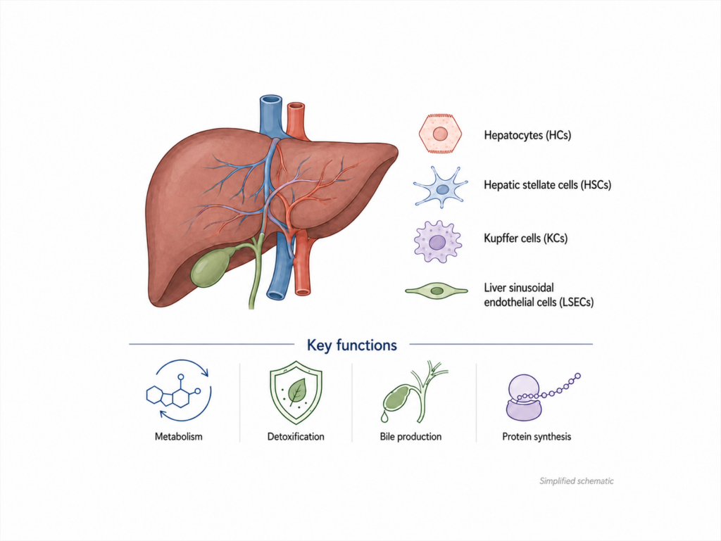
Hepatocytes are the predominant cell type in the liver. Together with other liver cells, such as hepatic stellate cells, Kupffer cells, and liver sinusoidal endothelial cells, they form a complex and highly specialized tissue environment.
Their main functions include the regulation of carbohydrate, lipid, and protein metabolism, detoxification of endogenous and foreign compounds, bile production, and the synthesis of plasma proteins such as albumin. These activities make hepatocytes essential for maintaining metabolic homeostasis.
In vitro, hepatocytes are used to study how the liver responds to drugs, toxic compounds, inflammation, metabolic stress, and disease-related conditions. Primary human hepatocytes are especially valuable because they can provide more physiologically relevant data than many immortalized liver cell lines.
Why hepatocyte culture is challenging
Although hepatocytes are extremely useful for research, they are also difficult to maintain in culture. Once removed from the liver and placed into standard 2D culture conditions, primary hepatocytes often begin to lose their mature liver-specific functions (2, 3).
This process is commonly described as dedifferentiation. It can involve changes in morphology, reduced metabolic activity, altered gene expression, and a decline in important liver functions such as albumin secretion, urea production, and cytochrome P450 enzyme activity (3).
For short-term experiments, traditional 2D hepatocyte cultures can still be useful. However, many toxicological and disease-related effects develop over several days or weeks. In these cases, the limited stability of 2D cultures becomes a major drawback (5, 7).
2D hepatocyte culture
Traditional hepatocyte culture is usually performed on flat plastic surfaces, often supported by extracellular matrix coatings such as collagen. Collagen sandwich cultures and Matrigel overlays have been used to improve hepatocyte polarity and function (2, 5).
These methods can help hepatocytes survive longer than in simple monolayer culture. However, they are not perfect. The use of biological matrices may introduce variability, and some culture conditions can influence hepatic gene expression. This can make it more difficult to interpret toxicology results, especially in long-term studies (5).
As a result, researchers have increasingly turned to 3D hepatocyte culture systems as an alternative.
3D hepatocyte spheroids
In 3D culture, hepatocytes are allowed to form compact spheroids instead of spreading as a flat monolayer. This arrangement better supports cell-cell interactions and can help maintain liver-specific functions for longer periods.
Primary hepatocyte spheroids have been shown to preserve important features of liver biology, including metabolic activity, hepatic gene expression, and functional markers such as albumin and urea production (7, 9, 10). Several studies have reported that 3D hepatocyte spheroids can remain viable and functional for several weeks, making them suitable for long-term toxicity testing (5, 7).
This is especially relevant for drug-induced liver injury studies. Some compounds do not show strong toxic effects in short-term assays but become harmful after repeated or prolonged exposure. A stable long-term hepatocyte model can therefore provide more meaningful information during drug development (5, 7).
Scaffold-free liver spheroids
One important approach in 3D hepatocyte culture is scaffold-free spheroid formation. In this system, cells are not embedded in an external gel or matrix. Instead, they self-assemble into spheroids, usually in ultra-low attachment plates.
This has several advantages. Scaffold-free systems are relatively simple to handle, compatible with high-throughput formats, and reduce the influence of undefined matrix components. They also allow researchers to generate spheroids in standard plate formats, such as 96-well plates.
However, the quality of the plate surface is critical. If the surface does not prevent cell attachment effectively, hepatocytes may spread across the well instead of forming a spheroid. If the well geometry or coating is inconsistent, spheroids may vary in size and shape. This can lead to poor reproducibility and less reliable assay data (12, 13).
For this reason, defined ultra-low attachment U-bottom plates are important for generating uniform liver spheroids (12, 13).
Why spheroid uniformity matters
In toxicology and drug screening, reproducibility is essential. Researchers need to know that differences in assay results are caused by the tested compound, not by variation in the model itself.
Uniform spheroids help reduce well-to-well variability. They also make imaging, viability assays, and functional readouts more consistent. Ideally, each well should contain one compact spheroid of similar size and morphology.
This is particularly important when working with primary hepatocytes, which can be more sensitive and variable than immortalized cell lines. Reliable spheroid formation can make experiments easier to compare across donors, species, compounds, and time points (7, 12, 13).
Applications of hepatocyte spheroids
3D hepatocyte spheroids are used in several areas of liver research.
One major application is drug-induced liver injury testing. Since hepatocyte spheroids can maintain liver-like functions over longer periods, they are useful for repeated-dose toxicity studies and for identifying compounds with delayed hepatotoxic effects (5, 7).
Another application is the study of cholestasis, where bile acid transport and bile-related toxicity are involved. Because 3D hepatocyte models can better preserve some liver-specific functions, they can be helpful for studying compounds with cholestatic liability (8).
Hepatocyte spheroids are also used in disease modeling. For example, human hepatic spheroids have been used to model steatosis and insulin resistance, which are relevant to fatty liver disease and metabolic disorders (11).
In addition, spheroids made from animal hepatocytes, such as dog or cynomolgus monkey hepatocytes, can support preclinical toxicology research. This is useful when researchers need to compare liver responses across species before moving into clinical studies (12, 13).
Human and animal hepatocyte spheroids
Primary human hepatocytes are highly relevant for predicting human liver responses. However, preclinical drug development often also involves animal models. Therefore, the ability to generate spheroids from different species is valuable.
Studies have shown that cryopreserved primary hepatocytes, including human and non-human primate hepatocytes, can be cultured in spheroid form under suitable conditions (13). Dog liver spheroids have also been explored for preclinical in vitro testing (12).
These models do not fully replace in vivo studies, but they can provide useful additional information. They may help reduce the number of unsuitable drug candidates earlier in development and improve the understanding of species-specific liver toxicity (12, 13).
The role of advanced 96-well plates
The development of improved 96-well spheroid plates has made 3D hepatocyte culture more accessible for routine laboratory work. A 96-well format allows researchers to test multiple compounds, concentrations, donors, or time points in parallel.
For hepatocyte spheroids, the plate should support efficient spheroid formation without unwanted cell attachment. It should also produce spheroids that are compact, round, and similar in size. These features are especially important when the model is used for toxicology assays, where small differences can affect the final interpretation (13).
Specialized polymer coatings can help create ultra-low attachment surfaces that promote reproducible spheroid formation. This is particularly useful for primary cells, which are often more difficult to culture than established cell lines (13).
Conclusion
Hepatocytes are essential cells for understanding liver function, drug metabolism, and liver toxicity. While traditional 2D cultures remain useful for some applications, they often lose liver-specific functions too quickly for long-term studies (2, 3, 5).
3D hepatocyte spheroids offer a more stable and physiologically relevant alternative. They support stronger cell-cell interactions, preserve important liver functions for longer periods, and are suitable for applications such as drug-induced liver injury testing, cholestasis research, fatty liver disease modeling, and preclinical toxicology (5, 7, 8, 11, 12).
For reliable results, however, the culture system itself matters. Defined scaffold-free conditions and high-quality ultra-low attachment plates can strongly influence spheroid quality, reproducibility, and assay performance (12, 13).
As liver research continues to move toward more predictive in vitro models, 3D hepatocyte spheroids are becoming an increasingly valuable tool for drug discovery, toxicology, and disease modeling.
References
- 1. Ding C., Li Y., Guo F., Jiang Y., Ying W., Li D., Yang D., Xia X., Liu W., Zhao Y., He Y., Li X., Sun W., Liu Q., Song L., Zhen B., Zhang P., Qian X., Qin J., He F. A Cell-type-resolved Liver Proteome. Molecular & Cellular Proteomics. 2016;15(10):3190–3202.
- 2. Kono Y., Yang S., Roberts E.A. Extended primary culture of human hepatocytes in a collagen gel sandwich system. In Vitro Cellular & Developmental Biology – Animal. 1997;33:467–472.
- 3. Elaut G., Henkens T., Papeleu P., et al. Molecular mechanisms underlying the dedifferentiation process of isolated hepatocytes and their cultures. Current Drug Metabolism. 2006;7:629–660.
- 4. Gómez-Lechón M.J., Tolosa L., Conde I., Donato M.T. Competency of different cell models to predict human hepatotoxic drugs. Expert Opinion on Drug Metabolism & Toxicology. 2014;10:1553–1568.
- 5. Bell C.C., Dankers A.C.A., Lauschke V.M., et al. Comparison of hepatic 2D sandwich cultures and 3D spheroids for long-term toxicity applications: a multicenter study. Toxicological Sciences. 2018;162:655–666.
- 6. Messner S., Agarkova I., Moritz W., Kelm J.M. Multi-cell type human liver microtissues for hepatotoxicity testing. Archives of Toxicology. 2013;87:209–213.
- 7. Bell C.C., Hendriks D.F.G., Moro S.M.L., et al. Characterization of primary human hepatocyte spheroids as a model system for drug-induced liver injury, liver function and disease. Scientific Reports. 2016;6:25187.
- 8. Hendriks D.F.G., Fredriksson Puigvert L., Messner S., Moritz W., Ingelman-Sundberg M. Hepatic 3D spheroid models for the detection and study of compounds with cholestatic liability. Scientific Reports. 2016;6:35434.
- 9. Bell C.C., Lauschke V.M., Vorrink S.U., et al. Transcriptional, functional, and mechanistic comparisons of stem cell-derived hepatocytes, HepaRG cells, and three-dimensional human hepatocyte spheroids as predictive in vitro systems for drug-induced liver injury. Drug Metabolism and Disposition. 2017;45:419–429.
- 10. Messner S., Agarkova I., Moritz W., Kelm J.M. Transcriptomic, proteomic, and functional long-term characterization of multicellular three-dimensional human liver microtissues. Applied In Vitro Toxicology. 2018;4:1–12.
- 11. Kozyra M., Johansson I., Nordling Å., Ullah S., Lauschke V.M., Ingelman-Sundberg M. Human hepatic 3D spheroids as a model for steatosis and insulin resistance. Scientific Reports. 2018;8:14297.
- 12. Raic A., Oberfrank C., Birk B., et al. 3D-Hunde-Lebersphäroide für die präklinische in vitro-Testung. BIOspektrum. 2021;27:736–738. https://doi.org/10.1007/s12268-021-1661-x
- 13. Ullrich A., Dieckhoff J., Schwartz V., Runge D., Hewitt P., Mentzel T. A novel polymer solution to generate ultra-low cell attachment surfaces and highly uniform spheroids in 3D primary cell cultures. 2021.